Frozen shoulder, also called adhesive capsulitis, is a thickening and tightening of the soft tissue capsule that surrounds the glenohumeral joint, the ball and socket joint of the shoulder. When the capsule becomes inflamed, scarring occurs and adhesions are formed. This scar formation greatly intrudes upon the space needed for movement inside the joint. Pain and severely limited motion often occur as the result of the tightening of capsular tissue.
There are two types of frozen shoulder: primary adhesive capsulitis and secondary adhesive capsulitis.
Primary adhesive capsulitis is a subject of much debate. The specific causes of this condition are not yet known. Diseases such as diabetes mellitus, and some cardiovascular and neurological disorders may also be contributing factors. In fact, patients with diabetes have a three times higher risk of developing adhesive capsulitis than the general population. Primary adhesive capsulitis may affect both shoulders (although this may not happen at the same time) and may be resistant to most forms of treatment.
Secondary (or acquired) adhesive capsulitis develops from a known cause, such as stiffness following a shoulder injury, surgery, or a prolonged period of immobilization.
With no treatment, the condition tends to last from two to three years. Many patients are unwilling to endure the pain and limitations of this problem while waiting for it to run its natural course. Even after many years, some patients will continue to have some stiffness, but no serious pain or functional limitations.
What are the signs and symptoms of frozen shoulder?
The major symptoms of frozen shoulder are pain and loss of motion.
The onset of symptoms may be gradual or sudden, depending on the cause of the condition. With primary adhesive capsulitis, the onset of symptoms is usually gradual. A sudden onset of symptoms may follow an injury to the shoulder.
The pain and loss of function associated with this condition can become so severe that it can significantly affect the quality of life, and prevent some patients from sleeping well or working.
How is frozen shoulder diagnosed?
The diagnosis of frozen shoulder is made only after a careful history and physical examination is performed. Pain and loss of motion can be symptoms of many shoulder conditions, so a detailed assessment of the shoulder's full range of motion is important. A history of surgery or injury, or the presence of illnesses such as diabetes, is information the physician needs in order to make the correct diagnosis.
It is important to recognize the different patterns of motion loss. Primary adhesive capsulitis is usually associated with loss of motion in all directions. Secondary adhesive capsulitis more often has more defined loss of motion; affecting some movements, but not others.
In most cases, the history and examination are sufficient to determine the presence or absence of frozen shoulder. Imaging may occasionally be necessary to confirm the diagnosis and to identify other underlying problems. X-rays cannot reveal the cause of shoulder stiffness in most cases of primary adhesive capsulitis. However, in secondary adhesive capsulitis, X-rays can show signs of arthritis, fractures, or metallic plates that may be contributing to motion loss.
How is frozen shoulder treated?
Non-Operative Treatment
For most patients with primary adhesive capsulitis, a supervised physiotherapy program will restore lost motion, although it can take more than six months to accomplish this. It is often necessary to combine a home program with supervised physiotherapy for maximum gains.
Shoulder stiffness that results from secondary adhesive capsulitis is generally more resistant to non-operative treatment. A supervised physiotherapy is always tried first. However, even an aggressive stretching program with an experienced physiotherapist is often ineffective when frozen shoulder follows an injury or previous surgery.
In many cases, non-steroidal anti-inflammatory medications can be very helpful with this condition. Other treatments such as ice, heat, and ultrasound may help alleviate some of the pain. These treatments are recommended as long as they are effective for the patient.
Supervised physiotherapy continues as long as the patient is making improvement. If after 12 to 16 weeks the patient is not improving or is actually getting worse, operative treatment should be considered.
Operative Treatment
Operative procedures to treat frozen shoulder include closed manipulation, as well as arthroscopic techniques. Operative treatment of primary adhesive capsulitis should only be considered once severe pain has subsided, and discomfort is present only at the extremes of motion. Severe pain represents the inflammatory stage of the disease. Surgery during this inflammatory phase may actually increase injury to the joint capsule, adding to the patient’s loss of motion.
Most patients who have not done well with a non-operative therapy program will do well with a closed manipulation or an arthroscopic capsular release procedure that is followed by aggressive motion therapy.
Closed manipulation is a technique in which the physician stretches the shoulder to break up the scar tissue and adhesions of the joint capsule. Although no incision is made, the patient is usually given a general and regional anesthetic that produces complete relaxation of the muscles.
·A patient who has had shoulder surgery within the past three months, or has a history of bone fragility (e.g. osteopenia), should not undergo closed manipulation due to the risk of damage to the soft tissue repair, nerve injury, or bone fracture.
·If there is a known cause of tightness outside of the joint (which may occur following some shoulder surgeries), an arthroscopic release is often necessary, and closed manipulation should not be attempted.
Pain management following manipulation is very important. If pain is not controlled, patients tend to limit shoulder motion, allowing scar tissue to form again. To minimize discomfort, long-acting analgesics are occasionally administered through a catheter. A comprehensive stretching program to restore lost motion is then started with a therapist. After the patient leaves the hospital, this program should continue until almost all motion has been recovered.
In some cases, a closed manipulation procedure may fail to restore motion to the shoulder. These patients may be candidates for selective arthroscopic capsular release, which has proven to be a safe, effective way to eliminate scar tissue from the capsule. During an arthroscopy, a small fiberoptic instrument is inserted into the joint. The scar tissue surrounding the joint is removed and a gentle manipulation follows. This will significantly reduce the risk of fracture or injury if the frozen shoulder has been present for some time. If necessary, other disorders within the shoulder can be addressed at the same time.
What types of complications may occur?
Complications after frozen shoulder surgery are generally infrequent. The most common problems associated with any of these procedures result from too little release, which fails to adequately reduce stiffness. Fractures of the humerus have been reported with closed manipulation. Older patients with fragile bones (osteoporosis) are more at risk for this type of complication. Although arthroscopic releases are relatively safe, releases in certain areas inside the joint have led to nerve injury.
What is the post-operative period like?
Following surgery:
·Patients usually remain in the hospital for one to two days. During this time, pain relief is provided either through a nerve block (sometimes with an indwelling catheter which provides a continuous interscalene block) or patient controlled analgesia.
·While in the hospital, patients begin an aggressive shoulder motion program supervised by the physiotherapist.
·Patients are encouraged to use the treated arm for daily activities. A sling is not worn.
·Patients are put on a home stretching program that is to be done between structured physiotherapy appointments.
·The strengthening phase of a rehabilitation program begins after the patient has achieved a full, pain-free arc of motion. This generally takes at least three months.
Impingement Syndrome & Rotator cuff tears
What is impingement syndrome?
Shoulder impingement syndrome occurs when the tendons of the rotator cuff and the subacromial bursa are pinched in the narrow space beneath the acromion (undersurface of the shoulder blade). This causes the tendons and bursa to become inflamed and swollen. This pinching is worse when the arm is raised away from the side of the body. Impingement may develop over time as a result of a minor injury, or as a result of repetitive motions that lead to inflammation in the bursa.
Particular shapes of the acromion may make certain individuals more susceptible to impingement problems between the acromion and the bursa. With age and the onset of arthritis, the acromion may develop bone spurs that further narrow this space. Impingement caused by bone spurs on the acromion is common in older patients who participate in sports or work activities that require overhead positions. Spurs may also result if one of the ligaments in the coracoacromial arch becomes calcified.
Impingement is classified in three grades:
·Grade I is marked by inflammation of the bursa and tendons.
·Grade II has progressive thickening and scarring of the bursa.
·Grade III occurs when rotator cuff degeneration and tears are evident.
What is a rotator cuff tear?
Continual irritation to the bursa and rotator cuff tendons can lead to deterioration and tearing of the rotator cuff tendons. The tendon of the supraspinatus muscle is the most commonly involved tendon among the rotator cuff muscles. It is subject to the most pinching of all the rotator cuff muscles.
Rotator cuff tears can be the result of a traumatic injury or deterioration over time. Symptoms may be present, but in many cases, the patient experiences no symptoms at all. In young active people, full thickness rotator cuff tears are fairly uncommon. When they do occur, they are usually the result of a high-energy injury to the rotator cuff that is associated with throwing or overhead sporting activities. In older people, rotator cuff tears tend to be the result of wear and tear over time. Several scientific studies have shown that up to 50% of the population at age 70 have rotator cuff tears; many of these people had no symptoms.
What are the signs and symptoms of impingement syndrome?
Most often the onset of symptoms is related to an episode of overuse. In many patients, the episode occurred some time in the past and the shoulder has failed to return to normal.
Impingement symptoms are marked by pain:
·The pain is sharp and intermittent in its early stages. As impingement progresses, the pain becomes more of a constant ache.
·Although pain is usually present after impingement sets in, the original event that led to the problem is often relatively minor and not remembered as painful.
·Once inflammation starts, simple movements may become painful. Overhead motions tend to increase the pain. There is less space for the bursa when the arm in this position, causing more compression on the bursa.
·Arm movements at waist level are not painful. In this position, there is more space for the bursa, and therefore it is less compressed.
·Pain usually increases at night for two reasons. First, inflammation and swelling tend to get worse as the shoulder is used during the day, and this can lead to more pain in the evening. Second, the mind is usually less occupied in the evening, allowing pain to become a major focus of attention.
What are the signs and symptoms of a rotator cuff tear?
The symptoms of a rotator cuff tear are very similar to those of impingement syndrome with the added complaint of weakness. This weakness will vary depending on which rotator cuff tendon has been torn. For example, if the supraspinatus muscle is involved (as is most often the case) weakness will be present with forward arm elevation and overhead activity. Many patients are at first unaware of how much strength they have lost when they tear the rotator cuff.
How are impingement and rotator cuff tears diagnosed?
With a careful history and physical examination, impingement and rotator cuff tears can be easily diagnosed. After the evaluation of symptoms, certain muscle tests will help to determine whether there are tears in the rotator cuff tendons and to rule out other conditions.
Further evaluation may include:
·A diagnostic injectioncan help us distinguish between impingement syndrome and a full thickness rotator cuff tear. A local anesthetic is injected into the inflamed bursae to eliminate the pain. If strength in the shoulder returns once the pain is blocked, it is likely that the weakness was due to pain and the rotator cuff tendons are not torn. A rotator cuff tear is suspected if strength does not return while the pain is blocked.
·X-rays can reveal signs of arthritis, fractures, and bone spurs on the acromion. They can also reveal changes in position of the humerus and scapula that may suggest a rotator cuff tear. These images are frequently negative in the early stages of injury since X-rays show bone structure but not soft tissue.
·An MRI (Magnetic Resonance Image) allows us to see muscle and other soft tissue not visible with X-ray.
·Ultrasound may also be used to diagnose a tear.
Will a torn rotator cuff require surgery?
This answer to this question depends on the condition of the other shoulder muscles and the age of the patient. Many older patients have no symptoms with a rotator cuff tear and continue to function without pain or disability. The goal of physical therapy is to maximize the function of the remaining tendons, and hopefully avoid surgery. In the younger age groups, particularly when tears are caused by a sudden injury, early surgery is generally recommended to insure a successful treatment outcome.
Generally speaking, a limited number (3) of steroid injections into the bursa are a safe, and often effective way to locally reduce inflammation and alleviate pain. These locally applied steroids do not have the same risks associated with the chronic use of oral steroids since the body does not systemically absorb them. However, it has been shown that repeated steroid injections can damage the quality of the rotator cuff tendons if a repair is later required.
I have been diagnosed with a rotator cuff tear in addition to frozen shoulder; why won’t the surgeon repair the rotator cuff now?
Surgery in the face of a frozen shoulder is not recommended because of the immobilization required after a rotator cuff repair. The shoulder becomes more inflamed after the surgery and the immobilization required to heal the cuff repair leads to increased stiffness. The only way to deal with this combination of problems is to allow physical therapy to stretch out the frozen shoulder. Once that has been accomplished, the rotator cuff repair can be performed. The shoulder will probably be stiffer than the average cuff repair after the immobilization period ends, but research has shown that physical therapy can help regain lost motion.
What is acromioplasty? How will it help my shoulder?
With acromioplasty, surgeons shave part of the acromion bone on the point of the shoulder. A ligament over the top of the shoulder is cut, and injured tissues are removed. This procedure is sometimes done to treat pinched tissues in the shoulder (called shoulder impingement). It is also used to treat tears in the rotator cuff. Acromioplasty can be done using an arthroscope. This slender instrument has a camera on the end that allows surgeons to work without making big incisions in the skin. Athroscopic acromioplasty is a less invasive procedure than open shoulder repairs, which require large incisions. For shoulder impingement and minor rotator cuff tears, acromioplasty has good results. Five years later, most patients have pain relief. They also have normal strength and motion in the shoulder. For rotator cuff tears, acromioplasty works best on minor tears and those on the undersurface of the tendon.
Instability, SLAP tears & biceps problems
If you first dislocated your shoulder in your teens what is the likelihood that recurrent shoulder instability will develop?
Research suggests that up to 80% of teenagers will develop recurrent instability after a first dislocation.
Should an open or arthroscopic surgery be performed?
The decision to have an open or arthroscopic repair depends on many factors. The cause of the instability, the total number of dislocations, and which technique the surgeon uses are important considerations when choosing the method of reconstruction. A thorough discussion with the surgeon of the available treatment options is essential. Regardless of the technique used, the rehabilitation following surgery is the same.
Why does my shoulder joint pop, crack and click?
Noises in the joints, such as popping, cracking or clicking, can be quite disturbing and cause concern. Often, these noises are not indicative of any underlying problem. Such noise often persists for years without any real problem developing. If there is no pain with cracks or clicks, you can generally assume it is being caused by the soft tissue in a joint. Noises that are associated with pain may indicate damage to the surfaces of the joint. Such cracks and clicks may be due to tears in labrum, which may snap over the other structures as the arm moves. If the labral tear is at the top of the shoulder it is called a SLAP tear. Sometimes the clicking may be due to the shoulder slipping in and out of joint. This is known as shoulder instability.
What is MDI?
MDI refers to a multidirectional laxity of the shoulder joint with associated instability. The instability generally results from stretching of the shoulder's supporting ligaments, which leads to increased movement of the glenohumeral joint. Research suggests that many patients (80%) will improve with physiotherapy alone. The patient's diligence and commitment to a daily physiotherapy programme is required for the best chance of success.
Will a labral tear heal without treatment?
There are no good natural history studies on labral injuries. There are acute (sudden) labral tear injuries that are likely to heal without surgery. In chronic (longstanding) cases, however, there are no successful non-operative treatments.
·a fall on the outstretched hand that drives the humerus upward and causes the superior labrum to tear.
·a sudden and often unexpected load applied to the biceps, which can cause a tear.
·extremes of external rotation and abduction (movement away from the body) during throwing that causes the labrum to "peel back" from its attachment.
Winging of scapula
What is Winging of the Scapula and what causes it?
The scapula (shoulder blade) is the largest bone of the shoulder complex and has the greatest number of muscles attached to it. These muscles both stabilise the arm to the body and move the arm around in space. All these muscles act at the same time at times and oppose each other at other times, but work together like a well trained team to allow the arm to move in space. If any of these muscles are not working in the right way at the right time this leads to a break in the rhythmic motion of the scapula. This is known as a scapula 'dyskinesia'. This leads to 'winging' of the scapula.
Winging of the scapula is a surprisingly common physical sign, but because it is often asymptomatic it receives little attention. However, symptoms of pain, weakness, or cosmetic deformity may demand attention.
Winging may be caused by injury or dysfunction of the muscles themselves or the nerves that supply the muscles.
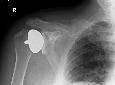
Shoulder arthritis and shoulder replacement
When is a Shoulder Replacement indicated for Arthritis of the Shoulder?
Most people with arthritis of the shoulder can manage with pain-killers, exercise and physiotherapy.
A shoulder replacement is considered if you have the following:
1. Severe pain which wakes you at night.
2. Pain which prevents you doing your daily activities.
3. Pain not controlled by pain-killers.
There are few contra-indications, if you have the above criteria. With modern techniques and implants, such as the Copeland surface replacement age is no longer a limiting factor.
What activities can I safely do after shoulder replacement?
The goal of shoulder arthroplasty is to relieve the pain from glenohumeral arthritis. It is unrealistic to expect to return to repetitive, heavy, overhead activities, which would put the replacement components at risk. Shoulder function after arthroplasty is also unlikely to allow the motions required by these activities.
Acceptable activities after a shoulder arthroplasty are:
·for those with previous experience in the activity: golf, shooting, and downhill skiing
Unacceptable activities are:
·football, gymnastics, hockey, rock climbing
·throwing sports
How painful is shoulder replacement surgery?
The anaesthetist takes great care to eliminate pain with appropriate analgesia both immediately after surgery and during the rehabilitation process. A long acting local anesthetic infused around the nerves of the joint is often used with general anaesthesia during surgery. These regional blocks will provide several hours of pain relief even after a patient has emerged from general anaesthesia. Alternatively a patient-controlled intravenous pump (PCA) is used in the early post-operative period for pain control. By the second or third day after surgery, oral pain relief medication is adequate through the early rehabilitation period (4-6 weeks).
How long before I can return to my normal activities after shoulder arthroplasty?
The time it takes to return to normal activity varies greatly from patient to patient. Most individuals have less pain at night or at rest in the first 2-4 weeks after surgery. Pain with activity persists longer, but generally decreases as the strength and function of the shoulder muscles improve. Full recovery usually takes 6-12 months.
I've heard that joint replacements sometimes "wear out" and need to be redone. What are the chances I may require a second shoulder arthroplasty?
Long-term studies show that 85-90% of total shoulder replacements are functioning well ten years after implantation, and 75-85% are doing well fifteen years after surgery. Over time, current advances in materials and techniques should improve these percentages even more.
What is the incidence of nerve damage in shoulder surgery?
Nerve damage following most common shoulder procedures is extremely rare. Large complex open procedures, such as revision shoulder replacements or complex fracture surgery are more likely to have a risk of significant nerve damage, but again this is not common.
What are the indications for a Reverse Geometry Shoulder Replacement?
The ball is fixed to the glenoid and the socket to the proximal end of the humerus in a reverse geometry shoulder replacement. This improves the mechanics of a cuff deficient shoulder joint. It is indicated in severe cuff arthropathy (shoulder arthritis in the presence of a massive irreparable rotator cuff tear) and salvage for failed shoulder replacements in the elderly.
Acromio-clavicular (AC) joint injuries
Do all AC separations require surgery?
No. In fact the vast majority of AC separations do very well with conservative treatment of the symptoms. Most AC injuries are grade I, II, or III and these generally do not require surgery. Usually the joint remains sore for two to six weeks and then full return to activity is the norm. Only unstable grade III injuries and high-energy AC separations, which are often the result of motor vehicle accidents, require surgery for full recovery.
Will the prominence over the AC joint ever go away?
The clavicle will become stable in its newly elevated position, but without surgery the "bump" will remain. The joint will function normally and will not remain tender to touch or movement. This minor cosmetic deformity will persist but will not interfere with overhead activities or participation in sports.
Are there downsides to a resection of the AC joint?
An AC resection is a procedure in which the AC joint (8mm) is removed through either an open or arthroscopic technique. Once the initial injury has healed and the clavicle has regained stability from scar tissue there is no functional loss with an AC resection. In the rare instance that the AC joint remains painful after a separation, but does not require stabilization, an AC resection is very effective in relieving pain without sacrificing function. If, however, the clavicle is unstable at the time of resection, a reconstruction procedure such as Weaver-Dunn is necessary to maintain the stability of the upper extremity.
Will I be able to return to athletics if an AC injury is not treated?
Absolutely. Most athletes in contact sports have had a low energy AC separation at some time in their careers. Except for the slight deformity that remains, there is no clinical significance to a healed AC separation. Occasionally high-energy AC separations that have disruption of the AC and CC ligaments will require surgery, but these injuries are usually apparent early on with a correct X-ray evaluation. Grade I, II, and most grade III AC separations will heal without treatment and a full return to sports can be expected.
I recently fell on the point of my shoulder and have been told I sprained my AC Joint. What is the best treatment and when is surgery indicated?
When a joint is first sprained, conservative treatment is certainly the best. Applying ice directly to the point of the shoulder is helpful to inhibit swelling and relieve pain. The arm can be supported with a sling which also relieves some of the weight from the shoulder. Gentle motion of the arm can be allowed to prevent stiffness, and early physiotherapy is often of benefit. Occasionally a steroid injection to AC joint may speed recovery if the injury is slow to settle. Most AC Joint injuries settle within 6 months, but a small proportion of patients continue to experience pain. This is usually because the small cartilage in the AC joint may have been torn and not healed. At this stage surgery is an option - in the form of an arthroscopic AC joint excision.
Success - Thank You
Your E-mail has been sent. We will be in contact with you soon.
Warning - Problem with your code
The security code you entered is incorrect. Please try again.
Fatal Error - Try again
SORRY
Your content caused an error and your email has not been sent.
Please remove any unusual characters and try again

)